Plankton as pictured by the Image in flow device of the CytoPro
Posted on
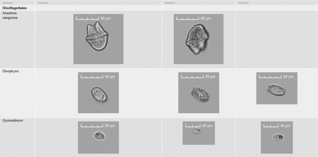
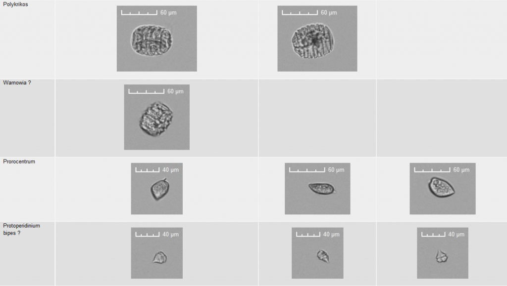
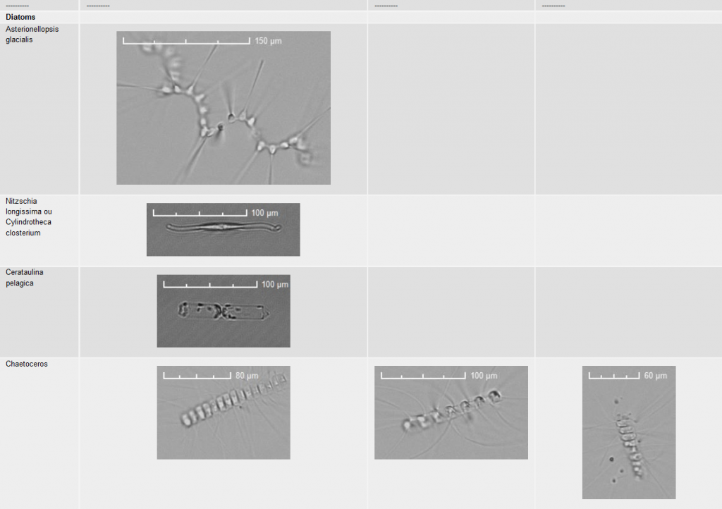
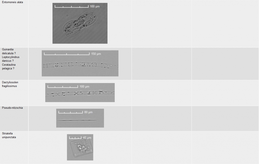
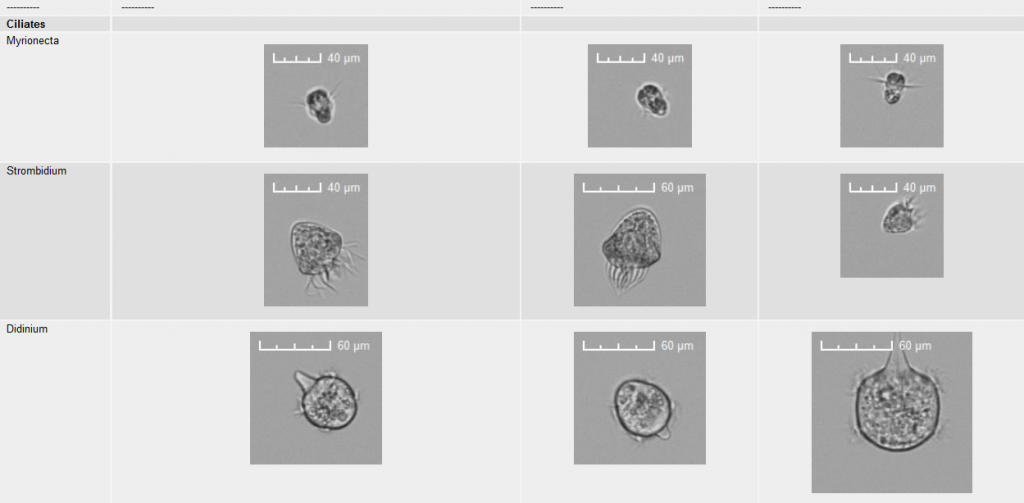
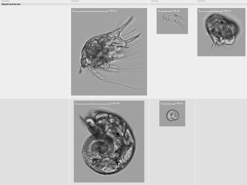
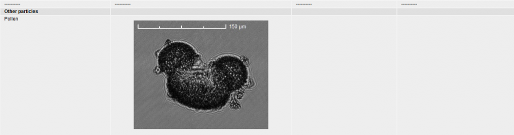
By implementing a camera into the flow cytometer, particles (cells) can be photographed as they flow. A group of particles is targeted (by drawing a region around it) and a limited amount of them can be photographed (up to 150 per analysis). This step allows for identification of the cluster forming cells defined by their optical properties (scatter and fluorescence intensities). The images are always coupled to the signals recorded as the particles cross the laser beam.
Here are some pictures taken by the IIF. Not only phytoplankton can be addressed. The capability of the Cytobuoy instruments in terms of size of the particles, volume analyzed (up to sevral mL), and imaging makes a big difference compared to more conventional flow cytometers.